Sleep and cells
Cellular defenses studied in sleep apnea research
Researchers studied sulforaphane responses in mice exposed to repeated low oxygen and in cultured neurons, examining NRF2, endoplasmic reticulum stress, and brain cell injury.

01
When oxygen falls and returns repeatedly
Obstructive sleep apnea repeatedly narrows or blocks the upper airway during sleep. When airflow is insufficient, blood oxygen falls, then recovers when breathing resumes.
Repeated oxygen shortage and reoxygenation can increase reactive oxygen species and damage proteins and lipids. Researchers model this as chronic intermittent hypoxia. Sulforaphane studies examine the signals by which brain cells respond to this stress.
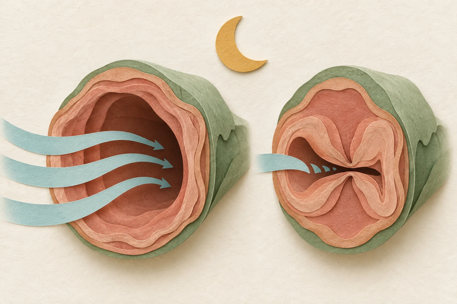
Airflow falls when the upper airway narrows. Concept illustration of repeated airway obstruction during sleep.
02
NRF2: how cells turn on defense genes
NRF2 regulates expression of defense genes in the nucleus. Under ordinary conditions, a system involving KEAP1 and CUL3 marks NRF2 for breakdown, keeping its level low. This regulation lets cells increase defense responses when needed.
Sulforaphane can affect KEAP1 sensing and inhibit marking NRF2 for breakdown. NRF2 can then accumulate and move to the nucleus, increasing expression of genes for antioxidant and detoxification enzymes. Zhang and colleagues identified this mechanism in cell experiments in 2004.
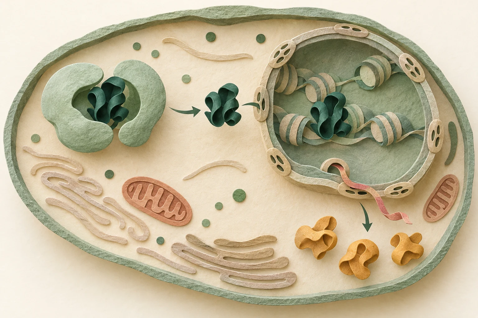
NRF2 that avoids breakdown can accumulate in the nucleus and regulate defense genes. Concept illustration of signaling from cytoplasm to nucleus.
Sources [5]
03
Brain responses in mice exposed to repeated low oxygen
In 2022, Li and colleagues exposed mice to intermittent hypoxia for four weeks and injected 0.5 mg/kg sulforaphane intraperitoneally each day before exposure. They measured defenses and injury in the hippocampus, a brain area involved in memory.
In the sulforaphane group, nuclear NRF2 and activity of the antioxidant enzyme SOD increased, while MDA, a lipid oxidation marker, decreased. Neuronal cell death fell and performance improved in a maze test of memory.
The researchers also measured autophagy related proteins including LC3 and Beclin-1 to examine antioxidant and cellular recycling responses.
Sources [1]
04
Endoplasmic reticulum: stress where proteins are folded
The endoplasmic reticulum folds and processes proteins. Misfolded proteins that accumulate there cause endoplasmic reticulum stress. When stress persists, PERK–ATF4–CHOP signaling can contribute to cell death.
In 2026, Zhong and colleagues studied rat derived PC12 cells, used as a neuronal model, and mice exposed to intermittent hypoxia. Sulforaphane treatment raised NRF2 and HO-1 defense responses while reducing endoplasmic reticulum stress signals and cell death markers. Mouse cognitive behavior tests also improved.
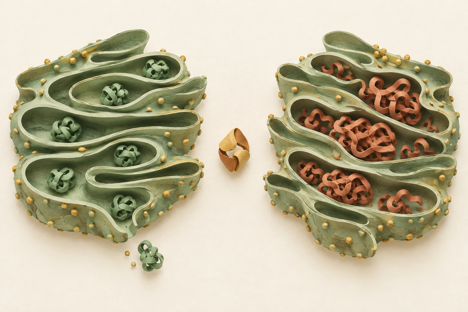
The endoplasmic reticulum folds and processes proteins. Concept illustration comparing organized processing with accumulation of misfolded proteins.
Sources [2]
05
Gene responses measured in the human airway
In 2009, Riedl and colleagues gave healthy adults a blended broccoli sprout preparation for three days. In cells sampled from the nasal mucosa, expression of defense enzyme genes including GSTM1, GSTP1, NQO1, and HO-1 increased.
The trial measured nasal defense gene expression; it did not assess sleep apnea symptoms.
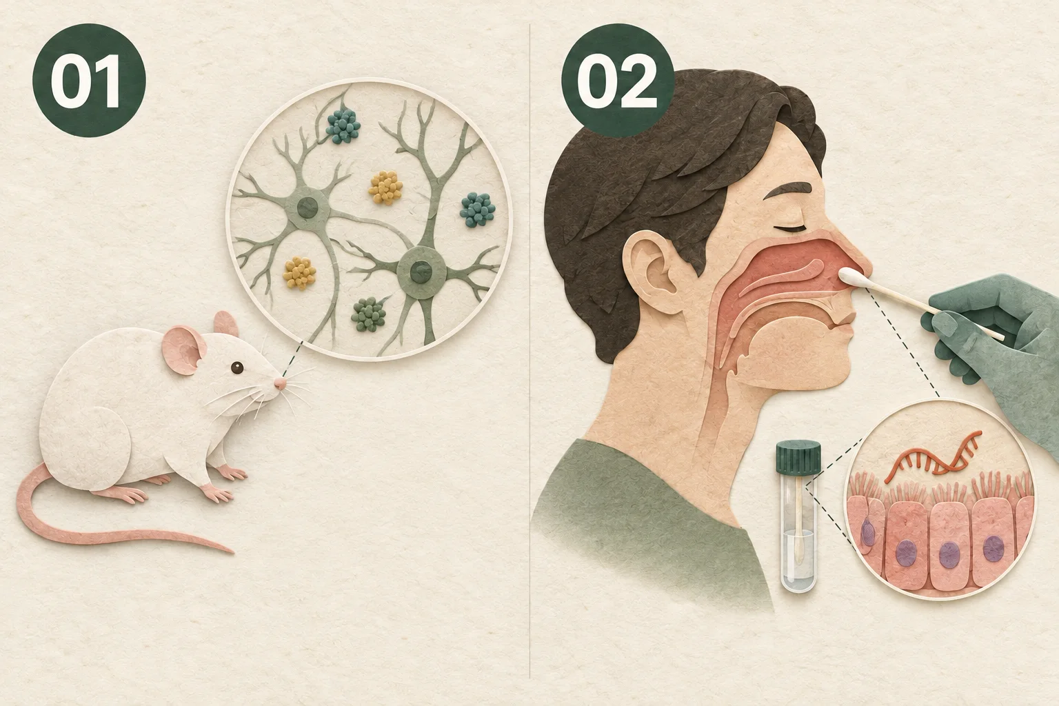
Changes studied in the hippocampus of intermittently hypoxic mice and the nasal mucosa of healthy adults.
- Mice · hippocampus
Antioxidant responses, injury markers, and memory behavior under intermittent hypoxia.
- Healthy adults · nasal mucosa
Defense enzyme gene expression after broccoli sprout intake.
References
Oct 19, 2026 sources reviewed
- Li et al. (2022) · Brain injury, NRF2, and autophagy in mice under intermittent hypoxia ↗
Mice exposed to intermittent hypoxia for four weeks received 0.5 mg/kg sulforaphane by intraperitoneal injection. Hippocampal NRF2, oxidative injury, autophagy markers, and cognition were assessed.
- Zhong et al. (2026) · Neuroprotective role of NRF2 activation against ER Stress-Mediated apoptosis in obstructive sleep apnea ↗
Study of endoplasmic reticulum stress and neuronal death in PC12 cells and mice.
- Riedl et al. (2009) · Oral sulforaphane increases Phase II antioxidant enzymes in the human upper airway ↗
Defense enzyme gene expression in nasal mucosa after broccoli sprout intake in healthy adults.
- US NHLBI · Causes of obstructive sleep apnea ↗
Public health explanation of upper airway narrowing and obstruction during sleep.
- Zhang et al. (2004) · Keap1 Is a Redox-Regulated Substrate Adaptor Protein for a Cul3-Dependent Ubiquitin Ligase Complex ↗
Cell study of KEAP1 and CUL3 marking NRF2 for breakdown and sulforaphane regulation.
Read next
Explore the Journal →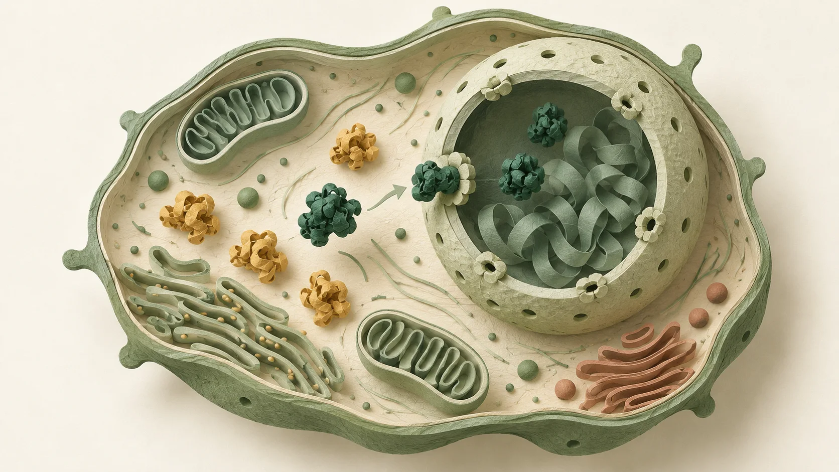
Cellular health basics
Sulforaphane and cellular health: how defense works
Explore how sulforaphane relates to cellular defense genes through KEAP1–NRF2 signaling, from reactive oxygen processing and antioxidant regeneration to human studies.

Research on cellular aging
Sulforaphane and cellular aging: reading the clocks
Explore cell senescence, immune cell responses after exercise, and gene expression based aging clocks through sulforaphane research.

SOD and antioxidant mechanisms
SOD, broccoli sprouts, and sulforaphane
Explore how SOD processes reactive oxygen and how sulforaphane relates to cellular defense genes, including enzyme formulations, human studies, and conversion in broccoli sprouts.

Aging well
Healthy aging: cognition and cellular defense
Explore sulforaphane and healthy aging through a 12 week cognition trial, a 42 month pilot study, and research on glutathione in blood immune cells.
Understanding cells
Cellular recycling: autophagy and sulforaphane
Autophagy encloses, breaks down, and reuses worn cell components. Explore cultured cell research linking sulforaphane with mTOR, TFEB, and ERK signaling.

Understanding ingredients
Phosphatidylserine and sulforaphane in cognitive health research
Phosphatidylserine helps organize cell membrane signaling, while sulforaphane regulates defense genes. Explore their roles and results from human cognition tests.