Understanding cells
Cellular recycling: autophagy and sulforaphane
Autophagy encloses, breaks down, and reuses worn cell components. Explore cultured cell research linking sulforaphane with mTOR, TFEB, and ERK signaling.
01
Enclose, break down, and reuse
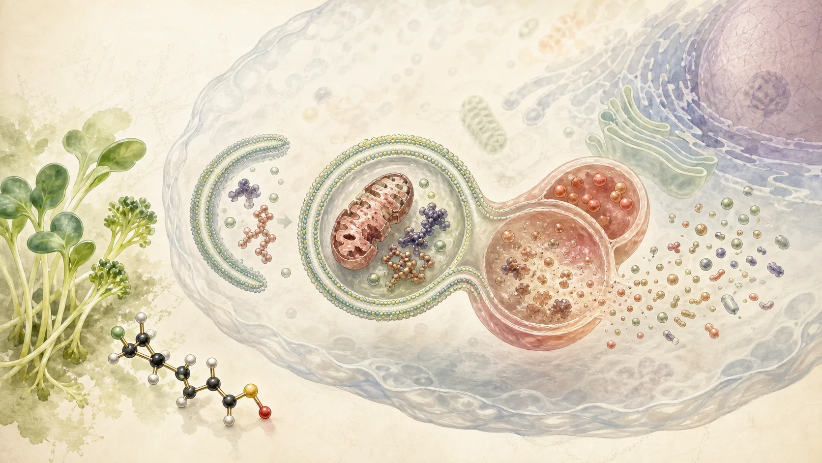
Cells break down damaged proteins and old internal structures for reuse. Autophagy is a process responsible for this recycling. Here we focus on macroautophagy, which encloses material in a double membrane and delivers it to a lysosome.
Lysosomes are small cellular compartments containing digestive enzymes. Autophagy collects and transports materials; lysosomes break them into forms the cell can use again.
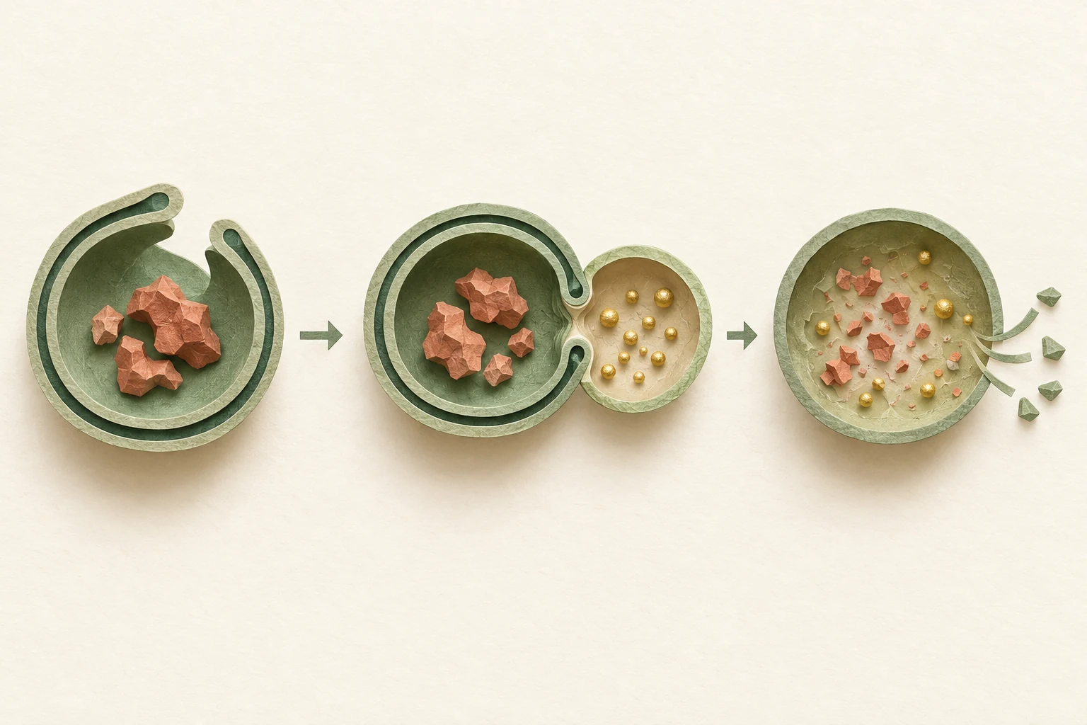
From left: enclosure, fusion with a lysosome, breakdown, and reuse. Concept illustration of autophagy.
- 01
Enclose with a double membrane
A membrane expands around the material. Once closed, it becomes a transport vesicle called an autophagosome.
- 02
Fuse with a lysosome
The autophagosome fuses with a lysosome, whose enzymes break down its proteins and cell structures.
- 03
Return usable materials
Smaller components return to the cytoplasm to build new structures or provide energy.
Sources [4]
02
mTOR: a signal regulating the start of recycling
Cells adjust synthesis and breakdown to nutrient conditions. Nutrient responsive mTORC1 supports growth and synthesis while suppressing the start of autophagy. When this signal falls, recycling can begin more readily.
In 2025, Plafker and colleagues treated cultured human retinal pigment epithelial cells (RPE-1) with 25 μM sulforaphane. Early insulin and mTORC2 signaling fell; by eight hours, phosphorylation of S6, a downstream mTORC1 marker, had decreased. Sestrin 2, which suppresses mTORC1, rose at the same time.
The researchers measured actual breakdown flow alongside signaling changes.
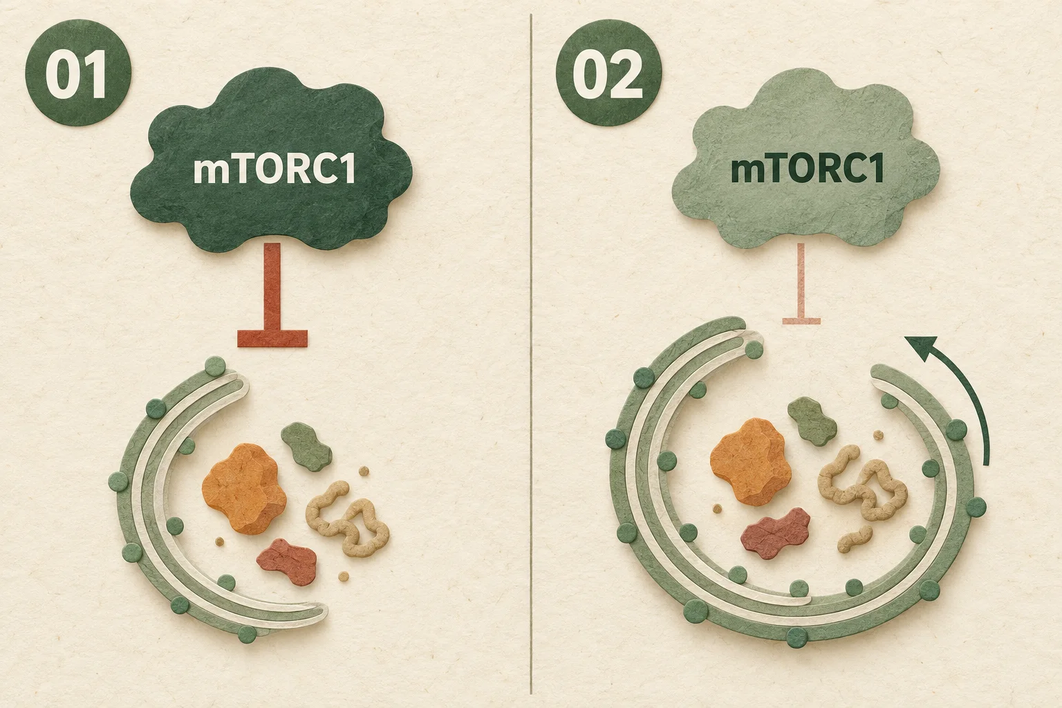
Lower mTORC1 signaling may ease suppression of autophagy initiation.
- Signal suppressing initiation
mTORC1 supports growth and synthesis while suppressing autophagy initiation.
- Eased suppression
With lower signaling, enclosure of material for recycling can begin more readily.
Sources [2]
03
TFEB: preparing the lysosomes that break material down
TFEB regulates expression of autophagy and lysosome genes in the nucleus. It coordinates genes needed to build and operate lysosomes.
A 2021 paper by Li and colleagues studied this pathway in human cervical cancer derived HeLa cells and other cells. A moderate rise in reactive oxygen species (ROS) after sulforaphane treatment was linked to calcium signaling. The calcium responsive enzyme calcineurin removed phosphate groups from TFEB, allowing TFEB to enter the nucleus and increase related gene expression.
In this experiment, TFEB activation followed a calcium signal independent of mTOR inhibition.
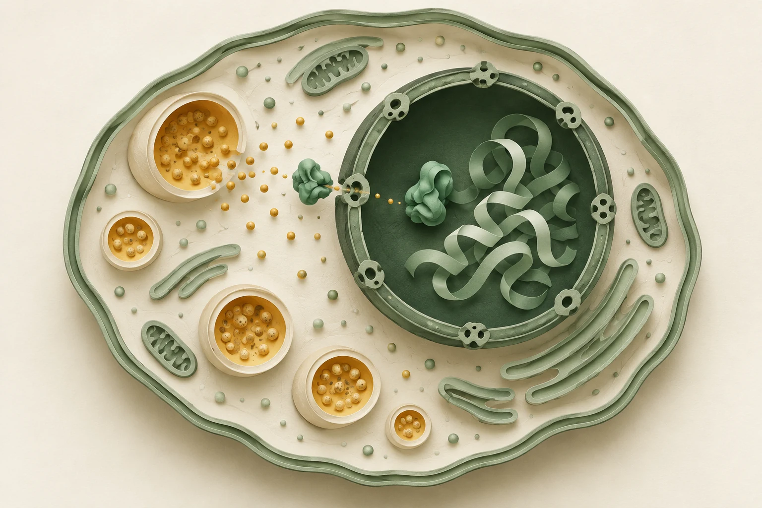
Calcium signaling near lysosomes is linked to TFEB movement into the nucleus, where it regulates autophagy and lysosome genes. Concept illustration.
Sources [3]
04
ERK: another link studied in neurons
In 2014, Jo and colleagues observed activated ERK signaling and increased LC3-II, a protein associated with autophagosome membranes, after sulforaphane treatment in cultured neurons. Pretreatment with the antioxidant NAC blocked both changes, suggesting a ROS–ERK route to autophagy.
Reducing or increasing NRF2 levels did not change the autophagy response. In these cultured neurons, the ROS–ERK pathway acted independently of NRF2.
Sources [1]
05
Measure breakdown flow, not only vesicle count
More autophagosomes or LC3-II can mean that new vesicles are forming faster or that later breakdown has stalled and vesicles are accumulating. The flow from formation to breakdown is called autophagic flux.
Plafker and colleagues compared GFP-LC3 signals with and without bafilomycin A1, which blocks breakdown. This difference estimated the material cleared and showed increased flux after sulforaphane treatment. After 24 hours, lysosome abundance and related gene expression also increased.
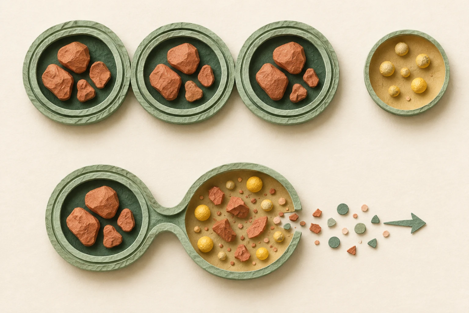
The upper path shows accumulated vesicles; the lower shows breakdown and reuse. Researchers compare with blocked breakdown to measure actual flux. Concept illustration.
Sources [2]
References
Oct 19, 2026 sources reviewed
- Jo et al. (2014) · Sulforaphane induces autophagy through ERK activation in neuronal cells ↗
Cultured neuron study of ROS–ERK signaling, LC3-II, and independence from NRF2.
- Plafker et al. (2025) · Sulforaphane acutely activates multiple starvation response pathways ↗
Human RPE-1 cells treated with 25 μM sulforaphane; mTOR signaling, flux measured with bafilomycin, and lysosome gene expression.
- Li et al. (2021) · Sulforaphane activates a lysosome-dependent transcriptional program to mitigate oxidative stress ↗
Cultured human cell study of TFEB nuclear movement through ROS, calcium, and calcineurin, plus lysosome gene expression. Published online in 2020 and in a 2021 issue.
- Nobel Prize Committee (2016) · Autophagy, breakdown, and recycling ↗
Official explanation of autophagosome formation, lysosome fusion, and recycling of cell components.
Read next
Explore the Journal →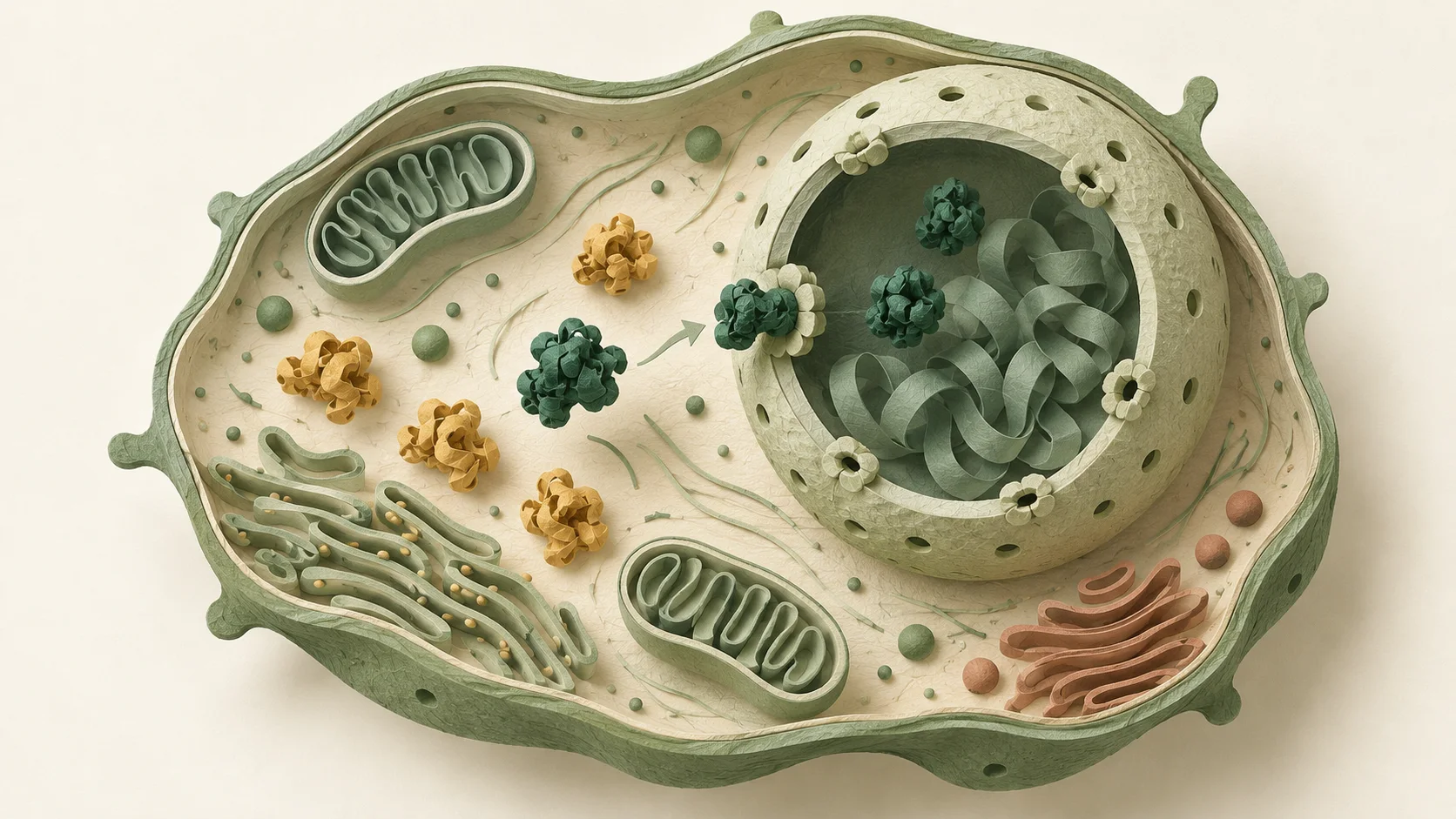
Cellular health basics
Sulforaphane and cellular health: how defense works
Explore how sulforaphane relates to cellular defense genes through KEAP1–NRF2 signaling, from reactive oxygen processing and antioxidant regeneration to human studies.
Research on cellular aging
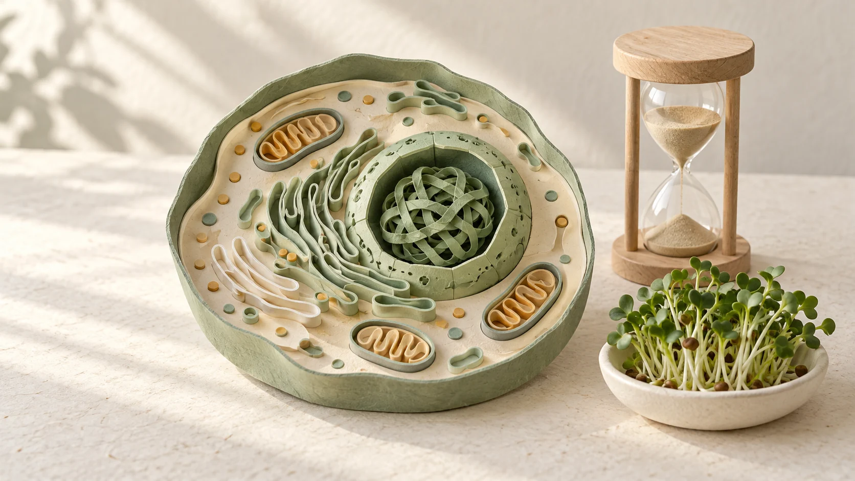
Sulforaphane and cellular aging: reading the clocks
Explore cell senescence, immune cell responses after exercise, and gene expression based aging clocks through sulforaphane research.

SOD and antioxidant mechanisms
SOD, broccoli sprouts, and sulforaphane
Explore how SOD processes reactive oxygen and how sulforaphane relates to cellular defense genes, including enzyme formulations, human studies, and conversion in broccoli sprouts.

Aging well
Healthy aging: cognition and cellular defense
Explore sulforaphane and healthy aging through a 12 week cognition trial, a 42 month pilot study, and research on glutathione in blood immune cells.
Sleep and cells
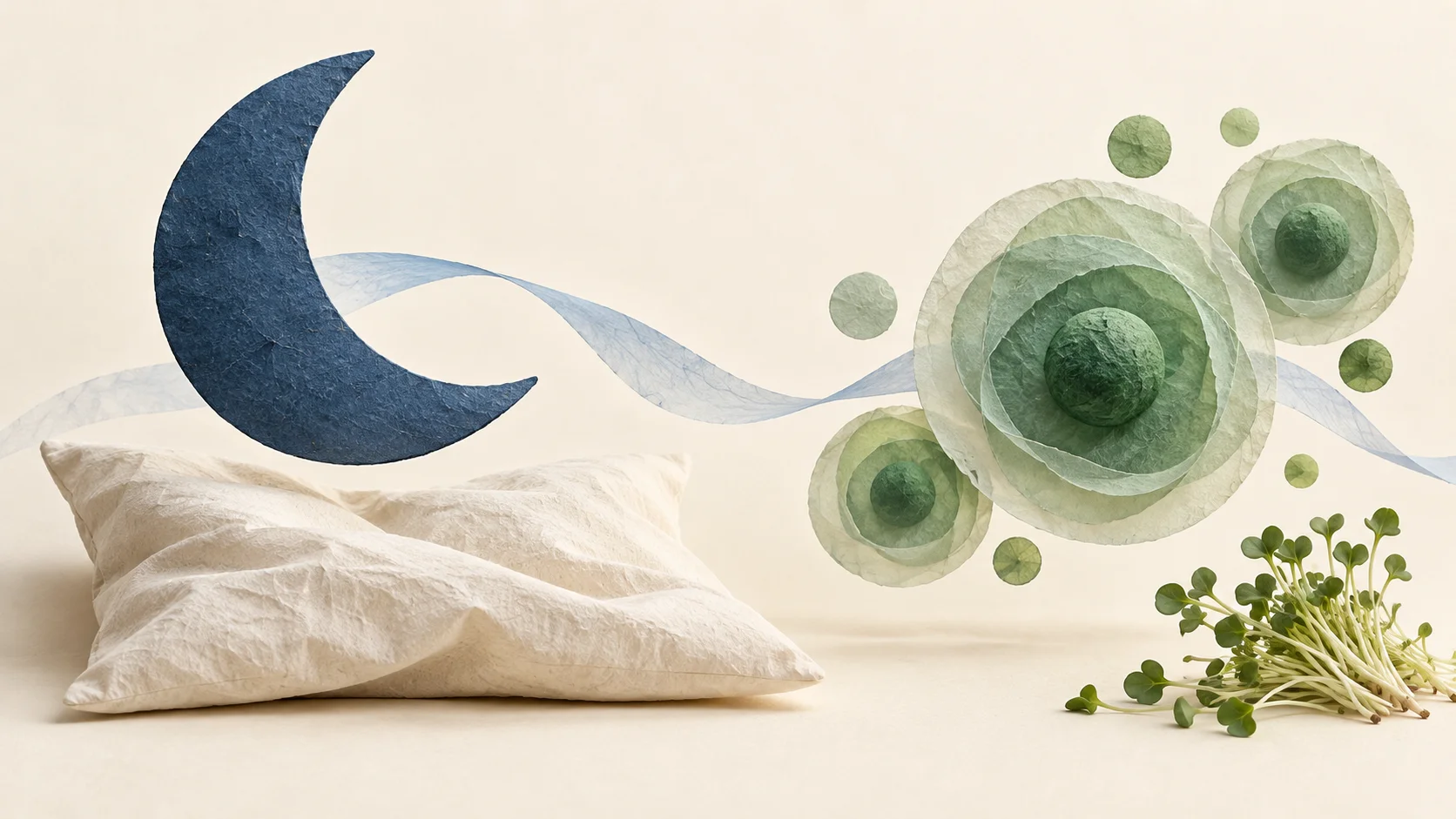
Cellular defenses studied in sleep apnea research
Researchers studied sulforaphane responses in mice exposed to repeated low oxygen and in cultured neurons, examining NRF2, endoplasmic reticulum stress, and brain cell injury.
Understanding ingredients
Phosphatidylserine and sulforaphane in cognitive health research
Phosphatidylserine helps organize cell membrane signaling, while sulforaphane regulates defense genes. Explore their roles and results from human cognition tests.